
Gastroenterology · August 23, 2026 · 12 min read
What Is ERCP? Uses, Benefits, Risks and Recovery Explained
By Dr. Vikas Sengar
Key takeaways
- ERCP is a specialized procedure combining endoscopy and X-rays to diagnose and treat diseases of the bile and pancreatic ducts.
- It is primarily used as a therapeutic tool to remove bile duct stones, bypass strictures, or relieve jaundice.
- The procedure is performed under sedation or general anesthesia, making it highly tolerable for patients.
- Common complications include pancreatitis, infection, and bleeding, which are managed carefully by a skilled specialist.
When dealing with disorders of the digestive system, standard imaging tests like abdominal ultrasounds, CT scans, or MRIs are excellent for visualization. However, when there is a blockage, narrowing, or stone within the drainage tubes of the liver, gallbladder, or pancreas, a more specialized intervention is required. This is where Endoscopic Retrograde Cholangiopancreatography, commonly known as ERCP, becomes essential. If you or a loved one has been advised to undergo this procedure, finding experienced care for an ERCP in Kanpur is crucial for ensuring a safe and effective treatment. In this comprehensive, patient-friendly guide, we will break down what the ERCP procedure involves, when it is considered, what benefits and risks it carries, and what to expect during your recovery under the care of a leading gastroenterologist in Kanpur.
What Is ERCP and What Does It Stand For?
Endoscopic Retrograde Cholangiopancreatography (ERCP) is an advanced medical procedure that combines upper gastrointestinal (GI) endoscopy with X-ray imaging (fluoroscopy). It is used to diagnose and, more importantly, treat conditions affecting the biliary tree (bile ducts) and the pancreatic ducts. The name breaks down as follows:
- Endoscopic: Refers to the use of an endoscope—a thin, flexible tube equipped with a light source and a camera—to view the inside of the digestive tract.
- Retrograde: Means "moving backward." During the procedure, the doctor injects a contrast dye backward into the ducts against the normal forward flow of bile and pancreatic juices.
- Cholangio: Refers to the bile ducts, which carry bile from the liver and gallbladder to the small intestine.
- Pancreato: Refers to the pancreatic duct, which drains digestive enzymes from the pancreas.
- Graphy: Refers to the recording of images, which are captured on X-ray screens during the injection of the contrast dye.
By merging endoscopic visualization with real-time X-ray monitoring, ERCP allows a specialist to not only identify obstructions but also perform therapeutic actions to resolve them immediately, saving many patients from the need for major open surgeries.
Why Is the ERCP Procedure Performed?
The primary function of the biliary and pancreatic ducts is to carry digestive juices into the duodenum, the first part of the small intestine. Bile, produced by the liver and stored in the gallbladder, helps digest fats. Pancreatic juice contains enzymes essential for breaking down proteins, carbohydrates, and fats. If these drainage tubes become blocked or narrowed, these juices back up, leading to symptoms like jaundice (yellowing of the skin and eyes), severe abdominal pain, and serious infections. An ERCP is performed primarily when there is a suspected blockage or structural problem in these ducts. It is important to note that while ERCP was once used as a purely diagnostic tool, non-invasive imaging technologies like Magnetic Resonance Cholangiopancreatography (MRCP) and Endoscopic Ultrasound (EUS) have largely replaced it for diagnostics. Today, the ERCP procedure is performed almost exclusively when therapeutic intervention (treatment) is planned during the same session.
What Conditions Can Be Treated with ERCP?
An ERCP may be considered for several conditions affecting the bile ducts, gallbladder, and pancreas:
Bile Duct Stones (Choledocholithiasis)
Gallstones can migrate from the gallbladder into the common bile duct, blocking the flow of bile. This is one of the most common reasons for performing an ERCP. Using specialized balloons or baskets, the doctor can perform an ERCP for bile duct stones, pulling the stones out into the small intestine where they can pass safely through the digestive tract.
Biliary and Pancreatic Strictures
Strictures are areas of abnormal narrowing in the ducts, which can be benign (caused by inflammation, infection, or prior surgery scars) or malignant (caused by tumors of the bile duct, pancreas, or gallbladder). During an ERCP, a gastroenterologist can place a small plastic or metal mesh tube (stent) across the narrowing to hold the duct open and restore normal drainage.
Acute Gallstone Pancreatitis
If a gallstone blocks the opening of the pancreatic duct, it can trap pancreatic enzymes inside the pancreas, causing severe, life-threatening inflammation (pancreatitis). Urgent ERCP to remove the obstructing stone can rapidly resolve the condition.
Bile Duct Leaks
A bile leak can occur as a complication of gallbladder surgery (cholecystectomy) or trauma. Placing a temporary stent during an ERCP helps redirect bile flow, allowing the leak to heal.
Tumors and Cancers
While ERCP does not cure cancers of the bile duct or pancreas, it is vital for palliative care, relieving obstructive jaundice and pain by placing stents to bypass tumors blocking the ducts. It also allows the doctor to take tissue samples (biopsies) or brush cytology for pathological testing.
How to Prepare for an ERCP
Proper preparation is critical for ensuring a safe procedure and clear imaging during an ERCP. Patients are given detailed instructions prior to their scheduled session:
- Fasting Guidelines: You must not eat or drink anything for at least 6 to 8 hours before the procedure. An empty stomach ensures that the endoscope can pass safely and reduces the risk of vomiting or inhaling stomach contents into the lungs (aspiration) under sedation.
- Medication Review: Inform your doctor about all medications you take. You may need to temporarily stop taking blood thinners (like aspirin, warfarin, or clopidogrel), nonsteroidal anti-inflammatory drugs (NSAIDs), or certain diabetic medications. Never stop taking prescribed medications without consulting your doctor.
- Allergies and Medical History: Tell your healthcare team if you have any allergies, particularly to iodine or contrast dyes used in X-rays, or if you have a pacemaker, kidney disease, or might be pregnant (as X-ray exposure must be avoided or minimized).
- Arranging Transport: Because you will receive sedation, you will not be allowed to drive or travel alone after the procedure. Arrange for a family member or friend to accompany you home.
What Happens During the ERCP Procedure?
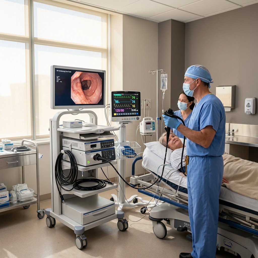
An ERCP is performed in a specialized endoscopy suite equipped with X-ray (fluoroscopy) machines. The procedure involves several structured steps:
Anesthesia and Patient Comfort
Before the procedure begins, you will receive local anesthetic spray to numb your throat and ease the passage of the scope. An intravenous (IV) line will be placed in your arm to administer sedatives. In many cases, deep sedation or general anesthesia is used to ensure you remain asleep, relaxed, and completely pain-free throughout the procedure.
Inserting the Duodenoscope
You will lie on your left side or stomach. The doctor will insert a specialized side-viewing endoscope (duodenoscope) through your mouth, down the esophagus, through the stomach, and into the duodenum. Air is gently pumped through the scope to inflate the tract and provide a clear view of the duodenal papilla (the Ampulla of Vater), which is the small opening where the bile and pancreatic ducts drain into the intestine.
Cannulation and Contrast Injection
The doctor passes a tiny plastic tube (catheter) through the endoscope and inserts it into the opening of the papilla. Through this catheter, a contrast dye is injected retrograde into the bile or pancreatic ducts. Real-time X-ray images (fluoroscopy) are taken immediately, highlighting the anatomy of the ducts on a screen and showing any stones, narrowings, or blockages.
Therapeutic Interventions
If a blockage or narrowing is identified, the doctor can perform immediate interventions through the scope:
- Sphincterotomy: Making a small cut in the muscle surrounding the opening of the bile duct (sphincter of Oddi) to widen it, facilitating stone removal or stent placement.
- Stone Extraction: Using inflatable balloons or wire baskets to pull stones out of the bile duct and sweep them into the intestine.
- Stent Placement: Inserting temporary plastic or permanent metal stents to bypass strictures and keep the ducts open.
- Biopsy: Taking tissue samples or cells from suspicious narrowings for laboratory analysis.
The entire procedure typically takes between 30 to 60 minutes, depending on the complexity of the treatment needed.
What to Expect During ERCP Recovery?
After the ERCP is complete, you will be monitored in a recovery room for 1 to 2 hours while the effects of the sedation wear off.
- Immediate Post-Procedure Care: You may feel slightly bloated or have mild abdominal gas cramps due to the air introduced during the procedure. A mild sore throat is also common and should resolve within 24 to 48 hours.
- Dietary Intake: In most cases, you will be allowed to drink liquids once your throat reflex returns, gradually progressing to a normal diet. If you had a complex procedure or have mild abdominal pain, you may be kept on a liquid diet or monitored in the hospital overnight.
- Activity Restrictions: You must rest for the remainder of the day. Avoid driving, operating machinery, or making important decisions for at least 24 hours. Most patients can return to normal daily activities within 2 to 3 days.
What Are the Possible Risks and Complications of ERCP?
ERCP is a highly effective procedure, but because it is invasive, it carries a higher risk of complications compared to standard upper endoscopies. It is not appropriate for every patient, and the benefits must be carefully weighed against the risks:
- Post-ERCP Pancreatitis (PEP): This is the most common complication, occurring in approximately 3% to 5% of patients. It is caused by irritation of the pancreas or pancreatic duct during cannulation. Most cases are mild, causing temporary back pain and vomiting that resolve with hospital monitoring, IV fluids, and pain management.
- Infection (Cholangitis): If a blocked bile duct is not fully drained, or if bacteria are introduced, it can lead to a serious infection. Antibiotics are often prescribed before and after the procedure to minimize this risk.
- Bleeding: Can occur at the site of a sphincterotomy, particularly if the patient is on blood thinners. Most bleeding is minor and stops on its own, but it can occasionally be controlled endoscopically during the procedure.
- Perforation: A rare but serious complication where a tear is made in the wall of the duodenum, esophagus, stomach, or bile duct. It may require surgical repair or endoscopic clipping.
- Adverse Reaction to Sedation: Breathing difficulties or heart rate fluctuations can occur but are closely monitored and managed by the anesthesia team.
When to Seek Immediate Medical Attention After ERCP
While minor discomfort is normal, you must contact your doctor or go to the nearest emergency department immediately if you experience any of the following warning signs:
- Severe, worsening abdominal pain that is not relieved by prescribed medication.
- High fever, chills, or sweating.
- Difficulty breathing or chest pain.
- Persistent nausea and vomiting, preventing you from keeping liquids down.
- Vomiting blood or material that looks like dark coffee grounds.
- Black, tarry, sticky stools (indicating internal bleeding).
- Dizziness, fainting, or severe weakness.
ERCP vs. Other Imaging Tests: How Do They Differ?
It is common for patients to wonder why they need an invasive ERCP when non-invasive tests are available. Here is a quick comparison:
- Ultrasound/CT Scan: Excellent first-line tests to detect gallstones or dilated bile ducts, but they cannot show small blockages in detail and cannot perform any treatment.
- MRCP (Magnetic Resonance Cholangiopancreatography): A specialized, non-invasive MRI scan that provides highly detailed 3D images of the bile and pancreatic ducts. It is purely diagnostic and carries zero risk of pancreatitis, but it cannot be used to remove stones or place stents.
- EUS (Endoscopic Ultrasound): Combines endoscopy with ultrasound to look at the walls of the GI tract and surrounding organs. It is highly accurate for diagnosing small stones and strictures, but has limited therapeutic capabilities compared to ERCP.
- ERCP: The only procedure that combines diagnostic imaging with immediate therapeutic intervention. If an MRCP or EUS confirms a stone or blockage, an ERCP is performed as the ERCP treatment to resolve the problem.
Role of Dr. Vikas Sengar for ERCP in Kanpur
Because ERCP is a complex, high-risk procedure, it requires a high degree of technical skill and experience. Dr. Vikas Sengar, a leading gastroenterologist in Kanpur, specializes in performing advanced therapeutic endoscopic procedures, including ERCP and SpyGlass cholangioscopy.
With a DM in Gastroenterology from SGPGI Lucknow and over 10 years of clinical experience, Dr. Sengar provides safe, precise, and patient-centered care at Medi Help Hospital, Sarvodaya Nagar, Kanpur. Equipped with modern fluoroscopy equipment and a dedicated endoscopy team, Dr. Sengar ensures that each patient undergoes a thorough evaluation to determine if ERCP is the most appropriate and safest option for their condition.
Frequently Asked Questions (FAQs)
Q1: Is an ERCP performed under general anesthesia?
An ERCP can be performed under deep conscious sedation or general anesthesia. Deep sedation makes you feel completely relaxed and asleep, while general anesthesia keeps you unconscious. The choice depends on the complexity of the procedure and patient health factors, ensuring a pain-free experience.
Q2: How long does it take to recover from an ERCP?
Most patients spend 1 to 2 hours in the recovery room and can go home the same day. You will need to rest for the remainder of the day. Mild sore throat or gas bloating may last 24 to 48 hours. Most people can return to light, normal activities within 2 to 3 days.
Q3: What is the most common complication of an ERCP?
The most common complication of ERCP is pancreatitis (inflammation of the pancreas), which occurs in approximately 3% to 5% of cases due to irritation of the pancreatic duct. It is usually mild and managed with temporary hospitalization, IV fluids, and pain relief.
Q4: Can bile duct stones pass on their own?
While very small stones may occasionally pass on their own, larger stones get stuck in the common bile duct. This block can cause severe pain, jaundice, and dangerous infections, making an ERCP procedure necessary to extract the stones safely.
Q5: Can I have an ERCP if I am pregnant?
Yes, but it is performed with extreme caution. Since ERCP uses X-rays, the procedure is only done during pregnancy if it is absolutely necessary (such as for severe cholangitis or pancreatitis) and cannot be delayed. Special shielding is used to protect the fetus from radiation.
Conclusion & Consultation Booking
An ERCP is a powerful, minimally invasive procedure that plays a critical role in treating complex disorders of the bile ducts and pancreas, saving many from open abdominal surgeries. However, because it carries specific risks, it must be performed by a highly qualified specialist. Book a consultation with Dr. Vikas Sengar for evaluation of digestive and liver-related concerns at Medi Help Hospital in Sarvodaya Nagar, Kanpur. To schedule your visit, call +91 98398 00199 or visit our online appointment booking portal.